
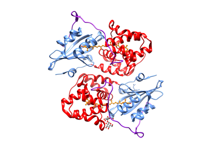
Ribbon view of the Orange Carotenoid Protein molecular structure (RCSB Protein Data Bank accession code 1M98). Key structural elements include the N-terminal domain (red), C-terminal domain (cyan), interdomain linker (purple), and the carotenoid chromophore (orange ball and sticks). The OCP crystallizes as a homodimer.
 Ribbon view of the Orange Carotenoid Protein by Ryan Leverenz and Cheryl Kerfeld is licensed under a Creative Commons Attribution-ShareAlike 4.0 International License. |
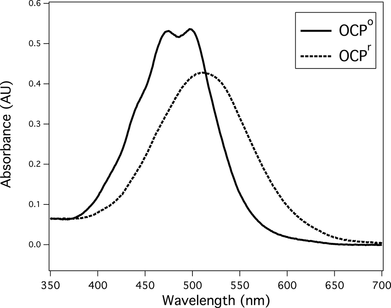
The visible absorbance spectrum of Orange Carotenoid Protein in its OCPo form (solid line) and illuminated OCPr form (dashed line). The OCPr form is obtained by illuminating the protein with a blue Light Emitting Diode.
Absorbance spectrum of Orange Carotenoid Protein by Ryan Leverenz and Cheryl Kerfeld is licensed under a Creative Commons Attribution-ShareAlike 4.0 International License. | ||
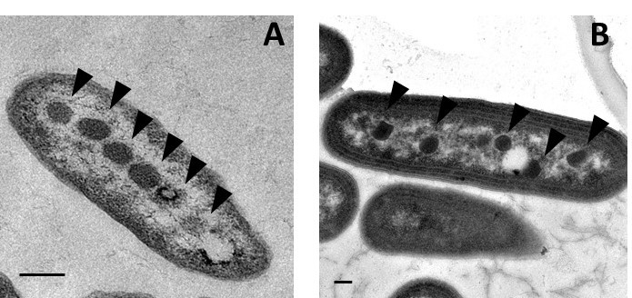
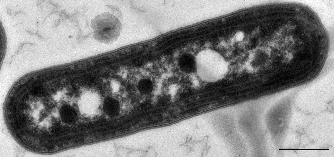
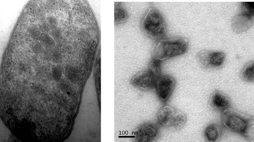
Electron micrograph of (A) alpha-carboxysomes in Halothiobacillus neapolitanus and (B) beta-carboxysomes in Synechococcus elongatus PCC 7942. Arrowheads highlight carboxysomes. Scale bars indicate 200 nm.
Electron micrograph of alpha and beta carboxysomes by Raul Gonzalez and Cheryl Kerfeld is licensed under a Creative Commons Attribution-ShareAlike 4.0 International License. |
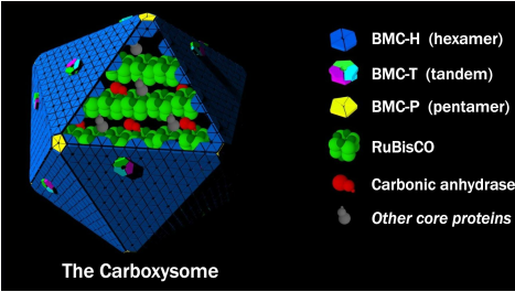
Model for the structure of the carboxysome. RuBisCO and carbonic anhydrase are arranged in an enzymatic core (organized by various core proteins) and encapsulated by a protein shell.
Model for the structure of the carboxysome by Raul Gonzalez, Seth Axen, and Cheryl Kerfeld is licensed under a Creative Commons Attribution-ShareAlike 4.0 International License. |
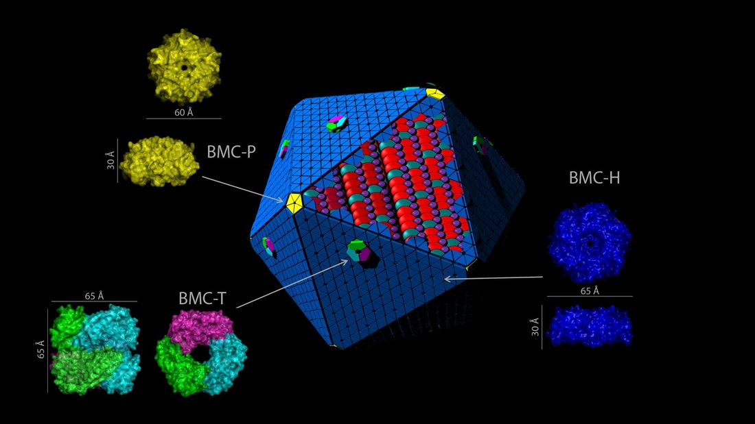
The three types of BMC shell proteins by Seth Axen, Markus Sutter, Sarah Newnham, Clement Aussignargues, and Cheryl Kerfeld is licensed under a Creative Commons Attribution-ShareAlike 4.0 International License. |
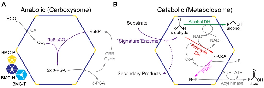
Generalized function schematic for experimentally characterized BMCs by Seth Axen and Cheryl Kerfeld is licensed under a Creative Commons Attribution-ShareAlike 4.0 International License. Based on a work at http://journals.plos.org/ploscompbiol/article?id=10.1371/journal.pcbi.1003898. |
Synechococcus elongatus PCC 7942 electron micrograph showing carboxysomes by Raul Gonzalez and Cheryl Kerfeld is licensed under a Creative Commons Attribution-ShareAlike 4.0 International License. |
PDU BMC genes expressed in Escherichia coli by Joshua Parsons, Steffanie Frank, Sarah Newnham, and Martin Warren is licensed under a Creative Commons Attribution-ShareAlike 4.0 International License. |